Description
Rehydrating/dehydrating agent of tissue and cytological samples
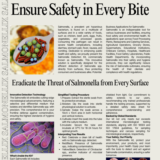
Introduction
Histology, cytology, and related scientific fields focus on examining the microscopic structure of tissues and cells. Achieving clear visualization of tissue and cellular structures requires precise sample processing. The histological sample processing involves several key steps, with three involving dehydration and subsequent rehydration. The initial step involves preparing the samples for infiltration, embedding them in paraffin, and then cutting the paraffin blocks into thin slices. In the second step, the samples are prepared for staining, and the final step
involves mounting the samples on glass slides. Since most embedding media, like commonly used paraffin, do not readily penetrate samples containing water, it is essential to perform dehydration first to facilitate the infiltration process. Once the samples are embedded in paraffin, cut into thin slices, and mounted on glass slides, they maintain their integrity for a specific period. However, before staining, it is necessary to remove the paraffin and rehydrate the sections. Only then can histological dyes be applied for staining. A similar procedure is followed for cytological samples, with dehydrating agents, primarily consisting of alcohols. One widely used dehydrating agent is denatured ethanol, which serves as the primary component in MenidiMedica Biotech Histanol. Histanol is a transparent, colorless, and flammable liquid known for its rapid action and high efficiency.
Product description
Other slides and reagents that may be used in staining:
– Fixatives such as MenidiMedica Biotech neutral buffered
formaldehyde solutions (Formaldehyde NB 10%)
– Dehydrating/rehydrating agent, such as MenidiMedica Biotech alcohol solutions
– Clearing agents, such as xylene or a substitute agent on the aliphatic hydrocarbons basis
– Infiltration and fitting agent, such as granulated paraffin
– High-quality glass slides for use in histopathology and cytology
– Differentiation agent, such as MenidiMedica Biotech Acid alcohol
– Bluing agents, such as MenidiMedica Biotech Scott’s solution or Bluing reagent
– Covering agents for microscopic sections and mounting cover glass, such as MenidiMedica Biotech Eukitt
– Cover glass, dimensions range from 18x18mm to 24x60mm
– Reagent for nuclear staining, such as MenidiMedica Biotech
Hematoxylin Harris
– Counterstaining reagents, such as MenidiMedica Biotech eosin solutions
Preparing histological sections for staining
– Fix the tissue sample tightly (10% NB Formaldehyde), rinse with water and dehydrate through series of ascending alcohol solutions (Histanol 100).
– Clear the sample with intermedium; in xylene or in a xylene substitute.
– Infiltrate and fit the sample in paraffin
– Cut the paraffin block to 4-6 µm slices and place them on a glass slide
Hematoxylin and eosin (HE) staining procedure, progressive
– Deparaffinize the section in xylene or in a xylene substitute – 3 exchanges, 2 min each
– Rehydrate using 100% alcohol (Histanol 100) – 2 exchanges, 5 and 3 min
– Rehydrate using 95% alcohol – 2 min
– Rehydrate in distilled water – 2 min
– Stain using Hematoxylin Harris – 3-5 minutes
Note: In the case of subsidence in the solution or a formation of metallic glow on the surface, reagent should be filtrated before use.
– Immerse the section in distilled or demineralized water until dye is no longer being released from the section
– Make nuclei turn blue using Scott’s solution or Bluing reagent – 1 min
Note: Finish the process of bluing after the nuclei turn blue If no Scott’s solution or Bluing reagent is available, rinse the sections under tap water for 3-5 minutes.
– Stain with one of eosin contrast solutions until the section is optimally stained – 15 seconds – 2 minutes
Note: Staining the sections in eosin alcoholic solutions causes intensive eosinophil color to show much faster (in under 15 seconds’ time). Recommended exposure time for eosin aqueous solutions is 90 seconds to 2 minutes
– Rinse under tap water – 2 min
– Dehydrate using 95% alcohol – 2 exchanges, 10-15 dips
– Dehydrate using 100% alcohol (Histanol 100) – 3 exchanges, 10-15 dips
– Clear the section in xylene or in a xylene substitute
Immediately after clearing apply an appropriate mount medium for covering/mounting on the section. If xylene was used, use one of mounting xylene-based media. If xylene substitute was used, use the appropriate covering agent for this case. Cover the section with a cover glass.
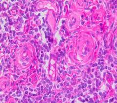
Result
Nucleus – dark blue
Cytoplasm, collagen, elastin, erythrocytes – various shades of pink (when staining with Eosin Contrast the shade is red-pink)
Note
Time periods of staining processes are not entirely standardized and they approximately correspond to clinical and laboratory practical experience. Intensity of staining depends on the period of immersion in the dye. Real staining protocol depends on personal requests and priorities.
Preparing the sample and diagnostics
Use only appropriate instruments for collecting and preparing the samples. Process the samples with modern technology and mark them clearly. Follow the manufacturer’s instructions for handling. In order to avoid mistakes, the staining procedure and diagnostics should only be conducted by authorized and qualified personnel. Use only microscope
according to standards of the medical diagnostic laboratory.
Safety at work and environmental protection
Handle the product in accordance with safety at work and
environmental protection guidelines. Used solutions and out of date solutions should be disposed of as special waste in accordance with national guidelines. Chemicals used in this procedure could pose danger to human health. Tested tissue specimens are potentially infectious. Necessary safety measures for protecting human health should be taken in accordance with good laboratory practice. Act in accordance with signs and warnings notices printed on the product’s label, as well as in MenidiMedica Biotech material safety data sheet.
Storing, stability and expiry date
Keep Histanol in a tightly closed original package at temperature between +15°C and +25°C. Keep in dry places, do not freeze and avoid exposing to direct sunlight. Date of manufacture and expiry date are Histanol 100 IFU ENG PDFprinted on the product’s label.